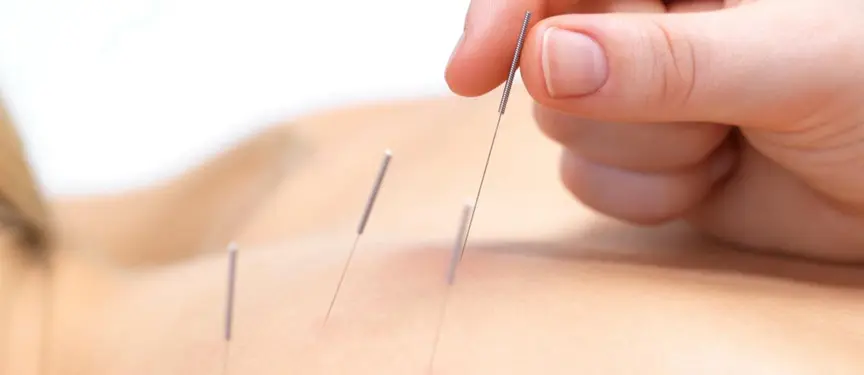
shoulder bursitis with chiropractor near me

Shoulder Bursitis: Causes, Complications, and Evolving Treatment Approaches

Understanding Shoulder Bursitis

Shoulder bursitis is an inflammatory condition affecting the bursa, small fluid-filled sacs that cushion and reduce friction between bones, tendons, and muscles in the shoulder. The subacromial bursa, situated beneath the top of the shoulder blade (acromion), is most commonly involved.

This condition can cause significant discomfort, often presenting with pain, tenderness, swelling, and restricted shoulder movement. If untreated, it may lead to compensatory changes in nearby joints or chronic conditions that are more difficult to manage.

Causes

Shoulder bursitis is usually caused by:

  1. Repetitive Movements: Overhead activities, such as painting or throwing, can irritate the bursa.
  2. Trauma or Injury: A direct blow or fall on the shoulder can cause inflammation.
  3. Chronic Conditions: Conditions like rheumatoid arthritis, gout, or diabetes may predispose individuals to bursitis.
  4. Poor Posture: Misalignment of the shoulder joint can increase pressure on the bursa.
  5. Rotator Cuff Dysfunction: Weakness or imbalance in the rotator cuff muscles can lead to improper joint mechanics, irritating the bursa.

Recent Insights into Shoulder Bursitis

1. Chronic Fibro-Adhesive Bursitis

Recent research has identified a subtype called fibro-adhesive bursitis. This condition, often linked to adhesive capsulitis (frozen shoulder), involves the formation of fibrous tissue within the bursa, which limits mobility and increases pain. Advanced imaging techniques like ultrasound are critical for its diagnosis, allowing for precise and targeted treatment.

2. Innovations in Physical Therapy

Physical therapy remains a cornerstone of shoulder bursitis management, emphasizing not only rehabilitation but also patient education. Adherence to prescribed exercise regimens is vital to prevent symptom recurrence. However, studies have highlighted a gap in patient satisfaction when therapy results are delayed or when negative past experiences diminish confidence. Individualized plans and clear communication are essential for improving outcomes.

3. Combining Treatment Approaches

Combining conservative treatments, such as corticosteroid injections, with structured physical therapy has shown greater efficacy in alleviating chronic bursitis symptoms compared to single modalities. This multi-faceted approach can improve both pain relief and shoulder function, particularly in complex or persistent cases.


Shoulder Bursitis and Nerve Impingement

While shoulder bursitis and nerve impingement are distinct conditions, they often intersect due to the close proximity of structures in the shoulder.

  • Swelling and Nerve Compression: Inflammation of the subacromial bursa can narrow the joint space, compressing nearby nerves like the suprascapular or axillary nerves. This may result in radiating pain, tingling, or weakness in the arm.
  • Coexisting Rotator Cuff Issues: Bursitis often co-occurs with rotator cuff injuries, further increasing the risk of nerve impingement.
  • Differentiating Symptoms: Bursitis and nerve impingement can present with overlapping symptoms, making precise diagnosis through imaging and clinical examination crucial.

Effective treatment that targets inflammation and improves joint mechanics can alleviate both bursitis and associated nerve symptoms.


Untreated Shoulder Bursitis: Impact on Surrounding Joints

When left untreated, shoulder bursitis may lead to compensatory changes in the cervical spine, elbow, and other parts of the upper kinetic chain.

1. Cervical Spine (Neck)

  • Increased Strain: Limited shoulder mobility can overburden the neck muscles, causing strain and discomfort.
  • Postural Imbalances: Chronic shoulder pain may lead to rounded shoulders or forward head posture, placing excessive stress on the cervical spine.
  • Secondary Conditions: Over time, these compensatory mechanisms may result in cervicogenic headaches or persistent muscle tension in the neck and upper back.

2. Elbow

  • Altered Mechanics: To avoid shoulder pain, individuals may rely on their elbow for movements, increasing strain on the joint.
  • Overuse Injuries: Conditions like tennis elbow or golfer’s elbow may develop due to repetitive compensatory use of the elbow.

3. Kinetic Chain Disruptions

The shoulder is part of a dynamic kinetic chain involving the neck, thoracic spine, and upper limbs. Dysfunction in the shoulder can create ripple effects throughout the chain, resulting in stiffness, abnormal movement patterns, and chronic discomfort.


Comprehensive Treatment Options for Shoulder Bursitis

Conservative Management

  • Rest and Activity Modification: Reducing repetitive movements to avoid further irritation.
  • Ice and Anti-inflammatory Medications: Effective for managing acute inflammation.
  • Physical Therapy: Focused on improving shoulder mechanics, restoring mobility, and strengthening muscles.

Chiropractic Care

Chiropractors address musculoskeletal imbalances that contribute to bursitis. Techniques may include joint adjustments, soft tissue therapies, and posture correction exercises to improve alignment and shoulder mechanics.

Advanced Medical Interventions

  • Corticosteroid Injections: Often combined with physical therapy for lasting symptom relief.
  • Aspiration: Draining excess fluid from the bursa to reduce swelling.
  • Surgery: Rarely required but may involve removing the bursa in severe or chronic cases.

Preventing Shoulder Bursitis and Its Complications

  1. Avoid repetitive overhead activities or take frequent breaks.
  2. Strengthen the rotator cuff and shoulder muscles with regular exercise.
  3. Address poor posture to minimize strain on the shoulder joint.
  4. Seek early treatment for shoulder pain to prevent chronic issues and compensatory injuries.

Conclusion

Shoulder bursitis, while common, is a manageable condition when diagnosed and treated early. Recent advancements in imaging and therapy highlight the importance of precise diagnosis and individualized treatment plans. By combining conservative care with patient-centered communication and addressing compensatory mechanisms, individuals can recover effectively and maintain optimal joint function.

Scheuermann Kyphosis with chiropractic care

Scheuermann Disease: Causes, Symptoms, and Treatment Options

Scheuermann Disease, also known as Scheuermann kyphosis, is a spinal condition that primarily affects adolescents. Characterized by a forward rounding of the back, it occurs due to an abnormal vertebral shape that leads to rigid hyperkyphosis. Unlike postural kyphosis, which is generally flexible and correctable with posture adjustments, Scheuermann Disease involves a structural deformity, making the curvature more rigid. This article provides an in-depth look at Scheuermann Disease, covering its causes, associated conditions, treatment options, and how chiropractic care can play a supportive role in managing symptoms.

What Causes Scheuermann Disease?

The exact cause of Scheuermann Disease remains unknown. However, it is believed to have a genetic component, as the condition often runs in families. Some researchers suggest that irregular growth of the vertebrae during adolescence may lead to the characteristic wedging shape of the bones. Another hypothesis is that microtrauma to the vertebrae during periods of rapid growth could result in inflammation and uneven vertebral development, though this theory is still under investigation.

Who is Most Affected by Scheuermann Disease?

Scheuermann Disease occurs more frequently in males than females, with most cases identified during adolescence. Growth spurts during this period appear to contribute to the development of abnormal vertebral shapes. The reason for the higher prevalence in males remains unclear but may relate to genetic factors or differences in skeletal development.

Symptoms Beyond Curvature

The most noticeable symptom of Scheuermann Disease is an exaggerated forward curvature of the thoracic spine, often creating a rounded or hunched appearance. Beyond this visible sign, other symptoms include:

  • Back Pain: Particularly common during periods of prolonged sitting or physical activity.
  • Muscle Fatigue: Especially in the back and lower limbs due to compensatory postural adjustments.
  • Reduced Flexibility: Limited motion in the thoracic spine, contributing to stiffness and discomfort.
  • Psychosocial Impact: Adolescents with visible deformities may experience self-esteem issues, affecting social interactions.

In severe cases, untreated Scheuermann Disease can lead to more significant health concerns, such as reduced lung capacity due to pressure on the thoracic cavity, although this is uncommon.

Conditions Associated with Scheuermann Disease

Scheuermann Disease is often linked with several other conditions and postural issues due to the altered biomechanics it causes:

  1. Scoliosis: Approximately 20–30% of individuals with Scheuermann Disease also present with scoliosis, a sideways curvature of the spine.
  2. Degenerative Disc Disease (DDD): Adults with Scheuermann Disease may be at a higher risk of developing degenerative disc disease due to uneven spinal loading, which can lead to stiffness and chronic back pain.
  3. Low Back Pain and Increased Lumbar Lordosis: To compensate for the excessive kyphotic curve in the thoracic spine, the lumbar spine often develops a more pronounced inward curve (lordosis), which can lead to chronic low back pain.
  4. Postural Imbalance and Muscle Tightness: Many patients experience muscle tightness, particularly in the hamstrings, hip flexors, and pectoral muscles. Back extensor muscles often become overstretched, increasing the risk of muscle strain and pain in the upper back, shoulders, and neck.
  5. Respiratory Issues in Severe Cases: In curvatures exceeding 70 degrees, lung compression may reduce breathing capacity, though this is rare and typically seen in untreated, severe cases.
  6. Neurological Symptoms (Rare): In advanced cases, nerve compression may occur, causing numbness, tingling, or weakness in the limbs.
  7. Psychosocial Challenges: Adolescents may experience social anxiety or depression due to the visible curvature, impacting self-confidence and quality of life.

Treatment Options for Scheuermann Disease

Treatments for Scheuermann Disease aim to reduce symptoms, improve posture, and prevent curvature progression, especially during growth years. While the structural vertebral shape cannot be fully reversed without surgery, there are several effective treatment options:

Conservative Management

  • Bracing: Bracing is often used in adolescents, particularly if the curvature is detected early during growth. A well-fitted brace can apply corrective pressure, slowing down or even slightly reducing the curve. However, bracing is only effective during growth periods and has minimal impact after skeletal maturity.
  • Physical Therapy: Physical therapy focuses on strengthening core and back muscles to improve spinal stability, flexibility, and posture. Exercises help reduce discomfort and muscle tension but do not change the vertebral shape.
  • Pain Management: Over-the-counter anti-inflammatory medications may help reduce pain, particularly after physical activity or prolonged sitting.

Surgical Intervention for Structural Correction

Surgery is reserved for severe cases with a curvature exceeding 70 degrees or when symptoms impair quality of life. Spinal fusion surgery is the most common approach, involving realignment of the vertebrae using rods, screws, or bone grafts. This procedure can significantly reduce the kyphotic curve and relieve severe symptoms, although it is a major procedure and considered only when other methods prove ineffective.

Chiropractic Care: Supporting Symptom Management

Chiropractic care plays a supportive role in managing Scheuermann Disease, focusing on symptom relief and enhancing spinal function rather than structural correction. While chiropractic adjustments cannot reverse the vertebral wedging, they can help alleviate pain and stiffness associated with the condition. Here’s how chiropractic care can assist:

  1. Spinal Mobilization: Gentle mobilization techniques improve flexibility and alleviate stiffness in the thoracic spine.
  2. Muscle Strengthening and Conditioning: Chiropractors work to strengthen surrounding muscles, providing better postural support and reducing muscle strain.
  3. Postural Education: Chiropractors provide guidance on posture, recommending specific exercises that promote thoracic extension and core strength.
  4. Pain Management: Manual adjustments, massage, and soft-tissue techniques relieve tension in muscles surrounding the kyphotic curve, reducing chronic pain.
  5. Exercise Prescription: Chiropractors may prescribe specific exercises aimed at improving spinal mobility, particularly in the thoracic region.

Long-term Prognosis for Scheuermann Disease

Once skeletal maturity is reached, the kyphotic curve tends to stabilize, meaning it doesn’t typically worsen in adulthood. However, if left untreated, Scheuermann Disease may lead to chronic back pain, reduced flexibility, and, in rare cases, respiratory complications. Regular physical activity, postural exercises, and core strengthening can help manage symptoms and prevent further complications.

What Happens Without Treatment?

If Scheuermann Disease is left untreated, several outcomes are possible, depending on the severity of the curvature:

  • Chronic Pain and Muscle Fatigue: Compensatory postural changes can cause chronic pain, particularly with prolonged sitting, standing, or physical activity.
  • Reduced Mobility and Flexibility: The thoracic spine may become progressively stiff, limiting the ability to bend or twist.
  • Psychosocial and Emotional Impact: Adolescents with visible curvature may experience social anxiety, body image issues, and depression. In adults, physical limitations or chronic pain can also impact mental health and quality of life.
  • Rare Respiratory Issues: Severe, untreated cases may lead to reduced lung capacity due to spinal compression of the thoracic cavity.

Scheuermann Disease, though a structural spinal condition, can be managed effectively with conservative treatments, lifestyle adjustments, and, in severe cases, surgery. Chiropractic care offers valuable support by relieving pain, improving flexibility, and enhancing quality of life. By understanding the long-term impact of Scheuermann Disease, individuals can make informed choices about treatment, helping to maintain their spinal health and well-being.


steroid injection

Steroid Injections: Uses, Benefits, Side Effects, and Alternatives

Have you ever suffered from neck, back, shoulder, or knee pain? After reviewing imaging results or if physical therapy has not been effective, your general practitioner might recommend a steroid injection. In some cases, your surgeon may suggest a steroid injection as an alternative to surgery. This article addresses frequently asked questions about steroid injections.

What is a Steroid Injection?

Steroid injections are a common treatment used to reduce inflammation and relieve pain in various musculoskeletal conditions. They are frequently administered for conditions like arthritis, tendinitis, and bursitis. These injections deliver corticosteroids directly into the affected area, providing targeted relief.

Why Are Steroid Injections Needed?

Steroid injections are mainly used to:

  1. Reduce Inflammation: Corticosteroids have strong anti-inflammatory properties that help control inflammation in joints, tendons, and muscles.
  2. Relieve Pain: By reducing inflammation, steroid injections can alleviate pain associated with chronic conditions like arthritis, allowing patients to regain mobility and comfort.
  3. Improve Mobility: Targeting painful, inflamed areas, steroid injections can improve the range of motion in affected joints, enhancing quality of life.
  4. Delay Surgery: For some patients, steroid injections may delay or even prevent the need for surgery, particularly in cases where inflammation is well-managed.

Who Can Perform Steroid Injections in Australia?

In Australia, qualified healthcare professionals with specialized training may administer steroid injections. These include:

  • General Practitioners (GPs): Many GPs with additional training can perform basic steroid injections for joint or soft tissue conditions.
  • Specialists: Orthopedic surgeons, rheumatologists, and sports medicine doctors often include steroid injections as part of their treatment plans.
  • Radiologists: For deeper or more complex injections, such as those around the spine, radiologists may use ultrasound or fluoroscopic guidance to ensure accurate placement.
  • Physiotherapists: Some physiotherapists, particularly those with advanced training and specific qualifications, may perform these injections under regulatory guidelines.

In certain cases, anesthetists may administer steroid injections, especially for precise pain management, such as spine injections.

How Are Steroid Injections Given?

Steroid injections are typically administered directly into the joint, tendon sheath, or muscle where inflammation is present. The procedure usually involves the following steps:

  1. Preparation: The area is sterilized to prevent infection. A local anesthetic may be applied to minimize discomfort.
  2. Guidance (if necessary): For complex injections, especially near the spine, ultrasound or fluoroscopy may be used to guide the needle precisely.
  3. Injection: The corticosteroid is injected, often with a local anesthetic for immediate pain relief.
  4. Aftercare: Patients are typically advised to avoid strenuous activities for a few days to allow the medication to take full effect.

What Are the Advantages of Steroid Injections?

  1. Targeted Relief: Unlike oral medications that affect the entire body, steroid injections offer localized relief focused on the affected area.
  2. Rapid Onset: Many patients experience pain relief within 24–48 hours after the injection, with effects lasting from several weeks to months, depending on the condition and individual response.
  3. Minimally Invasive: Compared to surgical options, steroid injections are less invasive, with fewer risks and a shorter recovery time.
  4. Reduced Dependence on Oral Medication: For those with chronic pain, steroid injections can decrease the need for oral pain relievers, which may carry systemic side effects over time.

How Frequently Can You Receive These Injections?

For many conditions, steroid injections are limited to three to four times per year in the same area to prevent potential complications associated with overuse. This interval helps maintain effectiveness while reducing the risk of side effects.

What Are the Potential Side Effects of Steroid Injections?

While generally safe under medical supervision, steroid injections may have potential side effects, especially with frequent use. Common side effects include:

  1. Pain and Swelling: Some patients experience a temporary “post-injection flare” with pain or swelling at the injection site.
  2. Skin and Soft Tissue Changes: Repeated injections can lead to skin thinning, pigment loss, or even muscle weakness near the injection site.
  3. Joint Infection: Though rare, infection is a risk with any injection, making sterile technique essential.
  4. Elevated Blood Sugar: Corticosteroids can temporarily raise blood glucose levels, important for patients with diabetes.
  5. Weakened Tendons or Ligaments: Frequent injections may weaken tendons over time, potentially increasing rupture risk.
  6. Bone Weakening: Repeated injections in the same location can cause localized osteoporosis or osteopenia, increasing fracture risk with minimal force.

Can Steroid Injections Cure the Underlying Condition?

Steroid injections are primarily a symptomatic treatment rather than a cure. They provide relief by reducing inflammation and pain but do not address the root causes of chronic conditions like arthritis or tendinitis. While they can improve mobility and quality of life, the underlying disease may continue to progress. Patients often combine steroid injections with physical therapy or lifestyle changes for more effective management.

Should Patients Try Conservative Treatments Like Chiropractic Care Before Steroid Injections?

Conservative treatments, such as chiropractic care, physiotherapy, and lifestyle modifications, are often recommended before resorting to steroid injections. These options can be beneficial, especially when strengthening and improving joint function can alleviate symptoms without medication.

For example, chiropractic care can help restore joint mobility, reduce muscle tension, and support spinal health, potentially reducing the frequency or necessity of steroid injections. Patients are encouraged to explore non-invasive treatments first, reserving steroid injections for cases where conservative approaches have not provided sufficient relief.

Conclusion

Steroid injections can be highly effective for managing pain and inflammation, but they should be part of a broader treatment plan. Conservative treatments are generally recommended before considering steroid injections. For those seeking quick pain relief, combining injections with physical therapy, lifestyle adjustments, and other supportive care can help maximize benefits while minimizing potential risks. Consulting a healthcare provider is essential to determine the suitability, frequency, and safety of steroid injections for each case.

Chiropractic management with Sacroiliac Joint Dysfunction

Sacroiliac Joint Dysfunction: Overview, Causes, and Chiropractic Care

Sacroiliac joint dysfunction (SIJD) refers to pain and improper functioning in one or both sacroiliac joints, located where the sacrum connects to the ilium of the pelvis. These joints play a key role in transmitting weight from the upper body to the legs and absorbing shock during movement. Dysfunction can arise when these joints become either too mobile (hypermobile) or too restricted (hypomobile), leading to discomfort and impaired movement.

Mechanisms and Causes of Sacroiliac Joint Dysfunction

Joint Instability(Hypermobility)

SIJ moves too much that causes the surrounding muscles and ligaments need to work harder to keep the joint stable.

Causes of hypermobility:

  • Trauma: Direct injuries or falls can misalign or damage the joint.
  • Pregnancy: Hormonal changes during pregnancy cause ligamentous laxity, increasing the risk of SIJD as the pelvis shifts.
  • Connective tissue disorders: Conditions like Ehlers-Danlos syndrome can lead to overly flexible joints, including the SI joint.

Joint Stiffness(Hypomoility)

In contrast, SI joint moves too little or becomes fixation that leads to compensatory movement patterns and pain.

Causes of hypomobility:

  • Arthritis: Inflammatory conditions such as osteoarthritis or ankylosing spondylitis may affect the sacroiliac joint, leading to dysfunction.
  • Postural imbalances: Poor posture, particularly in the lower back and pelvis, may lead to chronic stress on the SI joints.
  • Chronic inflammation: Repetitive stress or inflammation can cause fibrosis in the ligaments, reducing joint movement.
  • Prolonged immobility: Sedentary lifestyles or prolonged sitting can lead to stiffening of the joint.

Leg length discrepancies

A difference in leg length can place uneven stress on the SI joints, contributing to dysfunction. The joint on the shorter leg side may become hypermobile as it compensates, while the joint on the longer leg side may become hypomobile.

Muscle Imbalances

Muscles surrounding the pelvis, like the gluteal muscles and the hip flexors, play a significant role in stabilizing the SI joint. When the weakness and tightness occurring in these muscles, misalignment and uneven forces distribution in the SI joint can lead to dysfunction. Core weakness can put excessive strain on the SI joint, which fails to provide proper stabilization for the pelvis and lower back.

Repetitive Stress

Activities involving repetitive motions, like running, heavy lifting, or twisting, can cause wear and tear on the sacroiliac joint. Over time, this can lead to inflammation, joint degeneration, and dysfunction. Occupations that require heavy physical labor or athletes are at a higher risk for SIJD due to repetitive stress.

Inflammatory Component

The latest research has shown the inflammatory processes to play a role that develop chronic SIJD rather than pure mechanical causes. This emerging new concept suggests that individuals with autoimmune conditions such as ankylosing spondylitis (AS) or psoriatic arthritis (PS) may prone to SIJ inflammation.

Neurological Factors

A recent study found SIJ pain suffers often exhibit decreased proprioception in the pelvic region. It contributes to compensatory movement patterns and a higher risk of injury to lumbar spine and hip joints. Compared to the traditional strength and mobility exercises for SIJ, this focuses on and integrate proprioceptive training into rehabilitation programs.

Symptoms

The most common symptom of SIJD is pain, often described as sharp or aching, in the lower back or buttocks. This pain may radiate to the thighs or groin, and it often worsens with standing, sitting for prolonged periods, or climbing stairs. In some cases, individuals experience stiffness or reduced mobility in the lower back and pelvis.

Chiropractic Management of SIJD

Chiropractic care offers a non-invasive and effective approach to managing sacroiliac joint dysfunction. Chiropractors assess the alignment and mobility of the pelvis and spine to address the root cause of dysfunction. Several techniques may be employed:

  • Chiropractic Adjustments: Specific spinal manipulations are used to restore proper alignment and mobility to the SI joint. This can help alleviate pain and improve function by addressing joint restrictions or misalignments.
  • Soft Tissue Therapy: Chiropractic care may include soft tissue techniques such as massage or myofascial release to reduce muscle tension and inflammation around the SI joint.
  • Postural Correction and Exercises: Chiropractors often recommend corrective exercises to improve posture and strengthen the muscles supporting the pelvis, reducing the risk of future dysfunction.
  • Pelvic Blocking Techniques: Some chiropractors use specially designed blocks to adjust the pelvis and restore balance in the sacroiliac joints without the need for forceful manipulation.

Other Treatment Options:

Physical Therapy, Acupuncture, Anti-inflammatory drugs, Sacroiliac joint injections,Prolotherapy and platelet-rich plasma (PRP) therapy, Radiofrequency ablation(RFA), Bracing or SI belts and Surgery.

Precautions and Long-term Care

To prevent recurrences, individuals with SIJD should focus on improving core strength, flexibility, and posture. Chiropractors may recommend lifestyle adjustments, such as avoiding prolonged sitting or standing, using proper lifting techniques, and addressing any contributing factors like leg length discrepancies or poor footwear.

Conclusion

Sacroiliac joint dysfunction is a common condition that can significantly impact daily activities. Chiropractic care offers a holistic and patient-centered approach to managing SIJD, with a focus on restoring alignment, improving function, and preventing recurrence. Early intervention can lead to better outcomes and help patients regain their quality of life.

For those experiencing chronic lower back or pelvic pain, a comprehensive assessment by a chiropractor can determine whether sacroiliac joint dysfunction is the underlying cause and help develop a tailored treatment plan.

thumb pain with chiropractor near me

De Quervain’s Tenosynovitis: Symptoms and Treatment

Introduction

De Quervain’s tenosynovitis (also known as Mother’s thumb) is a painful condition that affects the tendons located on the thumb side of the wrist. It is caused by inflammation or degeneration of the sheath surrounding the tendons of the abductor pollicis longus (APL) and extensor pollicis brevis (EPB), which are responsible for thumb movement. The condition is most commonly associated with repetitive hand or wrist movements and can affect individuals who engage in activities involving gripping, pinching, or lifting.

Symptoms of De Quervain’s Tenosynovitis

The hallmark symptom of De Quervain’s tenosynovitis is pain and tenderness at the base of the thumb, which may extend to the lower part of the forearm. Other symptoms include:

  • Pain with thumb movement: Any movement that involves gripping, grasping, or pinching can exacerbate the pain.
  • Swelling near the base of the thumb: Swelling may be present along the wrist, and in some cases, a visible lump or thickening of the tendon sheath can occur.
  • Reduced range of motion: Pain and swelling may limit thumb and wrist mobility, making it difficult to perform everyday tasks.
  • A catching or snapping sensation: Some individuals report a sticking sensation when moving the thumb, caused by the inflamed tendons catching on the surrounding tissue.

Causes and Risk Factors

De Quervain’s tenosynovitis is primarily caused by overuse or repetitive strain on the tendons. Common activities that may lead to the condition include:

  • Repetitive gripping or pinching motions
  • Lifting a child or heavy objects
  • Participating in activities that involve wrist movement, such as tennis, gardening, or knitting

However, there are additional, lesser-known factors that may increase the likelihood of developing this condition:

  • Hormonal changes in women: Hormonal fluctuations, particularly during pregnancy and the postpartum period, can trigger De Quervain’s tenosynovitis. This condition is sometimes referred to as “mother’s thumb” or “mommy wrist” due to its frequent occurrence in new mothers. Hormonal changes, including increases in relaxin and estrogen, can lead to ligament laxity and make tendons more prone to injury and inflammation. As a result, women may develop this condition during pregnancy or postpartum.
  • Autoimmune conditions: There is a potential link between De Quervain’s tenosynovitis and autoimmune diseases such as rheumatoid arthritis. Inflammation from these conditions can increase the risk of tendon involvement.
  • Degenerative changes: Despite the “itis” suffix, not all cases of De Quervain’s involve inflammation. Research suggests that in some cases, degenerative changes in the tendons may be a factor, which could explain why anti-inflammatory treatments may not always be fully effective.

Gender Differences

Women are more prone to developing De Quervain’s tenosynovitis compared to men. Studies suggest that women are up to three times more likely to experience this condition. This higher prevalence is largely due to hormonal changes and the repetitive hand and wrist motions involved in tasks such as child care. In contrast, men typically develop this condition due to repetitive strain from manual labor or sports, without the influence of hormonal factors.

Diagnosis

mother's thumb test

De Quervain’s tenosynovitis is typically diagnosed based on a physical examination and medical history. One of the most common tests is the Finkelstein test, in which the patient makes a fist with the thumb tucked inside the fingers and bends the wrist toward the little finger. If this movement causes sharp pain on the thumb side of the wrist, it is a strong indication of De Quervain’s tenosynovitis.

Treatment Options

Treatment for De Quervain’s tenosynovitis focuses on reducing pain, improving function, and preventing recurrence. A combination of conservative and medical interventions is often recommended.

  1. Rest and Activity Modification
    Avoiding activities that aggravate the condition is essential for allowing the tendons to heal. Immobilizing the thumb and wrist with a splint may help prevent further irritation.
  2. Cold Therapy
    Applying ice to the affected area can help reduce swelling and numb the pain. It is advisable to apply ice for 15-20 minutes several times a day.
  3. Non-Steroidal Anti-Inflammatory Drugs (NSAIDs)
    Over-the-counter medications like ibuprofen or naproxen can help relieve pain and reduce inflammation.
  4. Physical Therapy
    A physical therapist may recommend exercises to strengthen the muscles around the thumb and wrist, improve range of motion, and reduce the risk of recurrence. Soft tissue therapy or manual techniques to release tension in the tendons may also be used.
  5. Corticosteroid Injections
    In cases where conservative treatments are not effective, corticosteroid injections may be administered to reduce inflammation. This treatment is generally very effective, with significant pain relief in most cases.
  6. Chiropractic Care
    Chiropractors can help with De Quervain’s tenosynovitis by using a variety of non-invasive techniques aimed at reducing pain, promoting tendon healing, and restoring function:

    • Joint Manipulation and Mobilization: Gentle adjustments to the wrist and thumb joints can help alleviate pressure on the tendons and improve mobility.
    • Soft Tissue Therapy: Techniques like myofascial release or trigger point therapy can reduce muscle tension and inflammation.
    • Cold and Heat Therapy: Chiropractors may use alternating cold to reduce inflammation and heat to improve blood circulation, speeding up recovery.
    • Kinesiology Taping: Applying kinesiology tape helps support the wrist and thumb, reducing strain and aiding in recovery.
    • Exercise Prescription: Chiropractors may recommend specific exercises to strengthen the muscles around the thumb and wrist.
    • Ergonomic Advice: Adjusting daily activities to avoid further tendon irritation is an essential part of chiropractic care.
  7. Surgery
    If symptoms persist despite conservative treatment, surgery may be recommended. The surgical procedure, known as tendon release, involves making a small incision to release the tendon sheath, reducing pressure and allowing the tendons to move freely. Surgery is typically considered only after other treatments have failed.

Prevention

Preventing De Quervain’s tenosynovitis involves minimizing repetitive thumb and wrist movements, especially in high-risk activities. Stretching exercises, ergonomic modifications, and regular breaks from repetitive tasks can all help reduce the risk. For individuals who have previously experienced the condition, continuing exercises to strengthen the wrist and thumb muscles may prevent recurrence.

Conclusion

De Quervain’s tenosynovitis is a common condition that can significantly impact hand and wrist function. Early diagnosis and treatment are essential for alleviating pain and restoring mobility. Conservative treatments are usually effective, but in severe cases, surgery may be required. Understanding the underlying causes, such as hormonal changes or autoimmune conditions, can help tailor treatment and prevention strategies for those at higher risk.

Carpal tunnel Syndrome with chiropractic care

Carpal Tunnel Syndrome (CTS):causes,symptoms and treatment

Carpal Tunnel Syndrome (CTS) is a common condition caused by compression of the median nerve as it passes through the carpal tunnel, a narrow passageway in the wrist formed by bones and ligaments. This compression can lead to pain, numbness, and weakness in the hand and wrist. Several factors contribute to the development of CTS, and new research has expanded our understanding of its causes, risk factors, and treatment options.

Symptoms of Carpal Tunnel Syndrome:

  • Pain or aching in the hand, wrist, or forearm.
  • Numbness or tingling, particularly in the thumb, index, middle, and ring fingers.
  • Weakness in the hand, leading to difficulty gripping objects or performing fine motor tasks.
  • Symptoms often worsen at night, sometimes disrupting sleep.

Causes and Risk Factors:

CTS is associated with repetitive motions that strain the wrist, such as typing or manual labor, but several other factors also play a role. These include:

  • Repetitive motions that involve prolonged or forceful hand and wrist movements.
  • Underlying health conditions, such as diabetes, hypothyroidism, or obesity, which increase inflammation.
  • Pregnancy and hormonal changes: Fluctuations in hormones, particularly estrogen and progesterone, increase susceptibility to CTS. These hormones can cause fluid retention and tissue swelling, exacerbating nerve compression. This explains the higher prevalence of CTS in females during pregnancy or menopause.
  • Arthritis: Rheumatoid arthritis (RA), in particular, can cause inflammation and swelling in the wrist, increasing the likelihood of median nerve compression. Osteoarthritis, although typically involving joint degradation, can also lead to structural changes in the wrist, potentially contributing to CTS.

CTS and Arthritis: Similarities and Differences

Similarities:

  • Both CTS and arthritis can cause pain, stiffness, and swelling in the wrist and hand.
  • Both conditions can be exacerbated by inflammation, whether from overuse, autoimmune responses, or structural damage.
  • Nerve involvement is common in both, as inflammation from arthritis or compression in CTS can lead to nerve pain and dysfunction.

Differences:

  • Carpal Tunnel Syndrome involves compression of the median nerve, resulting in numbness, tingling, and weakness in specific areas of the hand.
  • Rheumatoid Arthritis (RA) is an autoimmune disorder that leads to systemic joint inflammation, affecting multiple joints, including the wrists, with symptoms like joint swelling and deformities.
  • Osteoarthritis (OA) is characterized by the breakdown of cartilage in the joints, leading to pain and stiffness, but not directly causing nerve compression like in CTS.

Role of Vitamin D Deficiency in CTS:

Recent research has revealed a connection between vitamin D deficiency and more severe cases of CTS. Vitamin D is essential for reducing inflammation, and low levels can contribute to heightened nerve pain and tissue swelling. Studies suggest that supplementation may help manage inflammation and improve symptoms, though further research is needed to confirm the long-term benefits of this therapy.

Diagnosis:

CTS is diagnosed through physical exams and tests such as:

  • Tinel’s sign: Tapping the median nerve to elicit tingling.
  • Phalen’s test: Flexing the wrist for 60 seconds to provoke symptoms.
  • Ultrasound-guided diagnosis: The use of high-resolution ultrasound is becoming more common for diagnosing CTS. This method allows clear visualization of the median nerve and surrounding tissues, enabling more precise and individualized treatments, such as corticosteroid injections.

Treatment Options:

  1. Non-surgical treatments:

    • Rest and modification of activities that trigger symptoms.
    • Wrist splints, especially worn at night, to keep the wrist in a neutral position.
    • Non-steroidal anti-inflammatory drugs (NSAIDs) to reduce inflammation.
    • Acupuncture: Needling to reduce inflammation and pressure that compress the medial nerve.
    • Chiropractic care: Adjustments to relieve pressure on the median nerve.
    • Physical therapy: Includes stretching and strengthening exercises for the wrist.
    • Nerve Gliding Exercises: These exercises help the median nerve move more freely within the carpal tunnel, reducing pressure and improving symptoms. Studies show that regular practice of nerve gliding exercises can improve hand function and reduce pain.
  2. Steroid injections:

    • Corticosteroid injections can reduce inflammation and relieve symptoms. The use of ultrasound-guided injections ensures more accurate delivery of the treatment, targeting the affected area with greater precision.
  3. Surgical options:

    • Carpal tunnel release surgery may be necessary in severe cases.
    • Endoscopic carpal tunnel release offers faster recovery and less post-operative pain compared to traditional open surgery, but both approaches are effective long-term.
    • New surgical methods are being developed to minimize scarring and reduce recovery time, making surgery more efficient and less invasive.

Wearable Technology for CTS:

Recent advancements in wearable technology allow continuous monitoring of wrist movements and pressure. This technology helps identify patterns of repetitive strain that contribute to CTS. The data collected can be used to make personalized ergonomic recommendations, helping to prevent further nerve damage and manage early symptoms.

CTS and the Gut-Inflammation Connection:

Emerging research is exploring how gut health and inflammation are connected to CTS. Imbalances in gut microbiota may contribute to systemic inflammation, potentially exacerbating conditions like CTS. While further research is required, improving gut health through diet or probiotics may eventually become part of a holistic approach to managing inflammatory musculoskeletal conditions, including CTS.

Prevention:

  • Ergonomic adjustments to workstations, especially for those who use keyboards or perform repetitive manual tasks.
  • Frequent breaks to stretch and rest the wrists during repetitive activities.
  • Maintaining overall health through proper diet and exercise to reduce inflammation and manage underlying conditions such as obesity or diabetes.

Conclusion:

Carpal Tunnel Syndrome is a complex condition influenced by various factors, including repetitive motions, underlying health conditions, hormonal changes, and inflammation. Chiropractic care, non-invasive therapies like nerve gliding exercises, and newer diagnostic techniques such as ultrasound-guided treatments offer effective ways to manage symptoms. Understanding emerging research, such as the role of vitamin D deficiency, gut health, and wearable technology, further broadens the potential for personalized care and prevention strategies. By staying informed on these developments, individuals with CTS can explore a range of options for relief and recovery.

Chiropractic care for Tension headaches

Tension Headaches: Causes, Treatments, and Prevention Tips

Tension headaches are one of the most prevalent types of headaches, characterized by a dull, aching pain or pressure around the forehead, temples, or back of the head and neck. These headaches are typically caused by stress, poor posture, or muscle tension in the neck and upper back. Chiropractors provide effective treatment for tension headaches through spinal manipulation, lifestyle advice, and exercises to relieve muscle tension and correct posture.

Causes of Tension Headaches

Tension headaches primarily result from muscle tightness and strain in the neck, scalp, and upper back. These can be triggered by:

  • Stress: Emotional stress leads to muscle tension, especially in the shoulders, neck, and head.
  • Poor Posture: Prolonged sitting, slouching, or improper ergonomic setup at work can strain muscles and irritate joints.
  • Muscle Imbalance: Weakness in the muscles of the upper back and neck places additional strain on surrounding tissues.
  • Lack of Sleep: Sleep deprivation can cause muscle stiffness and tension, contributing to headaches.
  • Dehydration: Insufficient hydration can lead to muscle cramps and tension, triggering headaches.

Studies of Tension Headaches for further explanations

Impact of Poor posture and Muscle Imbalance 

Tension headaches, while often considered a benign condition, may actually have a significant link to musculoskeletal imbalances in the cervical spine and temporomandibular joint (TMJ) dysfunction. Misalignments in the upper cervical vertebrae can lead to referred pain patterns that mimic tension headaches. Recent studies have shown that myofascial trigger points in the suboccipital muscles and deep cervical flexors are frequently involved in tension headaches. These trigger points cause chronic tension in the scalp and neck, leading to persistent or recurrent headaches, especially during periods of stress or poor posture. Moreover, individuals with forward head posture are more prone to these headaches, as the added strain on the neck and upper back muscles contributes to muscle fatigue and spasms.

Impact of Sleep Disorders on Tension Headaches

Chronic tension-type headaches have been increasingly associated with sleep disturbances. A 2023 study published in Sleep Medicine found that poor sleep quality, including insomnia and obstructive sleep apnea, can exacerbate tension-type headaches. Disrupted sleep affects the brain’s ability to regulate pain, leading to increased sensitivity and headache frequency. Addressing sleep disorders in patients with tension-type headaches may significantly improve headache severity and frequency.

Emerging Research on Central Nervous System and Tension Headaches

Emerging research suggests that tension headaches might also be linked to disturbances in the central nervous system’s pain processing mechanisms. Chronic sufferers of tension headaches may develop a phenomenon known as central sensitization, where the brain becomes overly sensitive to pain signals, amplifying even mild stimuli into headache pain. This neural sensitivity may explain why some individuals experience frequent, daily headaches despite addressing external triggers.

Differentiating Tension Headaches from Other Types of Headaches

Tension headaches are often confused with other types of headaches, such as migraines or cluster headaches. However, there are distinct differences:

  • Tension Headaches: Characterized by a mild to moderate, steady ache or tightness around the head. The pain is typically bilateral (on both sides) and is not accompanied by visual disturbances or nausea, which are common with migraines.
  • Migraines: Migraines are typically more intense, with throbbing pain that often occurs on one side of the head. Migraines are often accompanied by nausea, vomiting, and sensitivity to light or sound.
  • Cluster Headaches: These are rare but extremely painful headaches that occur in cyclical patterns or clusters. The pain is typically focused around one eye and can last for weeks or months.

Chiropractic Techniques for Tension Headaches

Chiropractors employ a range of techniques to relieve tension headaches:

  • Spinal Manipulation: Misalignments in the cervical spine (neck region) can cause muscle tension and contribute to headaches. Chiropractic adjustments restore proper alignment, reducing pressure on nerves and relieving headache symptoms.
  • Postural Corrections: Chiropractors help correct posture through specific exercises and ergonomic recommendations, addressing one of the primary causes of tension headaches.
  • Soft Tissue Therapy: Massage and trigger point therapy target tight muscles in the neck, shoulders, and upper back, relieving tension that often leads to headaches.
  • Ergonomic Advice: A chiropractor may assess your workspace setup and recommend ergonomic adjustments to reduce strain on the neck and shoulders, which can help prevent tension headaches in the long term.

  • Exercise Prescription: Strengthening exercises for the neck, shoulders, and upper back can help build endurance and reduce muscle tension, which can mitigate the triggers of tension headaches.

What Can Be Done to Improve or Prevent Tension Headaches

Lifestyle changes and self-care strategies can significantly reduce the frequency and intensity of tension headaches. Some helpful tips include:

  • Maintain Good Posture: Avoid slouching or hunching over screens. Keep your computer screen at eye level and sit with your shoulders relaxed.
  • Take Frequent Breaks: If you spend long hours working at a desk, take breaks to stretch your neck, shoulders, and upper back every 30 minutes to an hour.
  • Stay Hydrated: Drink plenty of water throughout the day to prevent dehydration, which can trigger muscle tension and headaches.
  • Manage Stress: Practice relaxation techniques such as deep breathing, meditation, or yoga to reduce stress.
  • Sleep Well: Ensure you get enough sleep, as lack of rest can increase muscle tension and headache occurrence.
  • Exercise Regularly: Physical activity helps reduce muscle tension and improve circulation, both of which can prevent tension headaches.

Who Is Prone to Tension Headaches?

Certain groups of people are more likely to experience tension headaches. These include:

  • People with Sedentary Lifestyles: Individuals who sit for long periods, particularly those with poor posture, are at greater risk due to muscle tension from inactivity.
  • Stress-Prone Individuals: Those experiencing high levels of emotional or physical stress are more likely to develop tension headaches.
  • Office Workers: People who work at computers for long hours, particularly in poorly ergonomic environments, are prone to tension headaches due to posture-related muscle strain.
  • People with Muscle Imbalances: Weakness or tightness in neck and upper back muscles can predispose individuals to tension headaches, as the imbalance increases strain on supporting structures.

Conclusion

Chiropractic care provides an effective, non-invasive approach for treating tension headaches by addressing their root causes, such as spinal misalignments and muscle tension. Through spinal adjustments, postural correction, and lifestyle advice, chiropractors help reduce headache frequency and severity. Additionally, addressing lifestyle factors such as posture, hydration, stress, and sleep quality can further reduce the likelihood of developing tension headaches.

TMJ Chiropractor helps Lock Jaw

How to Fix Lockjaw: Causes, Symptoms and Treatments

Introduction

Lockjaw refers to the inability to open the jaw fully, which can be caused by different conditions, notably tetanus and temporomandibular joint (TMJ) dysfunction. Understanding these conditions helps in distinguishing their causes, symptoms, and treatment options.

1. Tetanus: A Rare Cause of Lockjaw

Tetanus is a bacterial infection caused by Clostridium tetani, which produces toxins affecting the nervous system, leading to severe muscle contractions, including those of the jaw, known as trismus or lockjaw. Although rare in developed countries due to widespread vaccination, it remains a serious condition.

Symptoms of Tetanus Include:

  • Painful muscle spasms, especially in the jaw and neck.
  • Difficulty swallowing or breathing.
  • Fever, sweating, and increased heart rate.

Treatment: Tetanus requires immediate medical intervention, including antitoxins, antibiotics, muscle relaxants, and vaccination if the patient isn’t already immunized.

2. TMJ Dysfunction: A More Common Cause of Lockjaw

TMJ dysfunction involves issues with the temporomandibular joint, which connects the jaw to the skull. This dysfunction can lead to pain, limited jaw movement, and in severe cases, lockjaw. Common causes include injury, arthritis, and poor posture.

Symptoms of TMJ Dysfunction Include:

  • Pain or tenderness in the jaw.
  • Clicking or popping sounds when opening or closing the mouth.
  • Difficulty chewing or opening the mouth wide.
  • Headaches, earaches, or neck pain.

3. Posture and Its Impact on TMJ Function

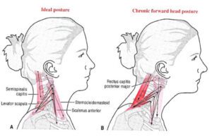
Poor posture, especially forward head posture, can strain the muscles around the neck and jaw, worsening TMJ dysfunction. Slouching or keeping the head forward can misalign the jaw, leading to increased pressure on the TMJ, contributing to muscle tension and joint inflammation.

How Posture Affects TMJ:

1. Forward Head Posture and Increased Strain

TMJ mucles

Forward head posture occurs when the head is positioned too far in front of the body, often as a result of prolonged computer use, texting, or slouching. This posture shifts the balance of the head, placing additional strain on the muscles of the neck, shoulders, and jaw. The altered positioning can create tension in the muscles that control the TMJ, leading to:

  • Jaw misalignment: The position of the jaw may shift, causing the TMJ to function improperly.
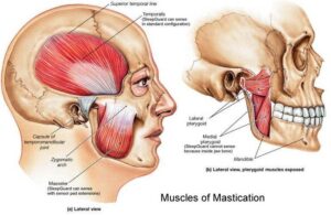
  • Increased muscle tension: The muscles involved in jaw movement, such as the masseter, temporalis, and pterygoid muscles, can become overworked, leading to pain and dysfunction.

2. Impact on the Occlusion and Bite

Bad posture, particularly involving neck misalignment, can alter the way the upper and lower teeth come together (occlusion). This can cause:

  • Abnormal bite pressure: Uneven bite forces can result in uneven wear on the teeth and excessive strain on the TMJ, contributing to pain and discomfort.
  • Malocclusion (misaligned bite): Prolonged bad posture may exacerbate or contribute to malocclusion, further affecting TMJ function.

3. Increased Joint Compression

TMJ articular Disc

Poor posture can lead to increased compression within the TMJ itself. When the head is positioned forward, the lower jaw (mandible) is often forced into a retruded (backward) position. This can result in:

  • Pressure on the TMJ: The backward displacement of the jaw may increase pressure on the joints, leading to inflammation, pain, and restricted movement.
  • Cartilage wear: Over time, this increased compression can contribute to degenerative changes in the TMJ, such as cartilage wear or displacement of the articular disc.

4. Neck and Shoulder Imbalance

Slouching or rounded shoulders contribute to muscle imbalances in the upper back, neck, and shoulders, affecting TMJ function indirectly by:

  • Reducing mobility: Tight muscles in the neck and shoulders can limit movement in the jaw, leading to restricted range of motion in the TMJ.
  • Tension headaches and neck pain: Bad posture can cause or worsen tension headaches and neck pain, often overlapping with TMJ-related symptoms, making it harder to pinpoint the source of discomfort.

5. Chronic Pain and TMD

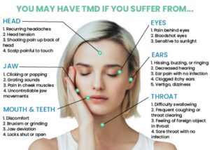
Chronic bad posture can perpetuate a cycle of pain and dysfunction. Over time, poor alignment and muscular strain may lead to temporomandibular disorders (TMD), which are characterized by symptoms such as:

    • Jaw pain or tenderness
    • Clicking, popping, or locking of the jaw
    • Difficulty chewing or speaking
    • Earaches, headaches, and facial pain

4. Chiropractic Care and TMJ Dysfunction

Chiropractic care can be highly effective in managing TMJ dysfunction by addressing the underlying musculoskeletal issues contributing to the condition.

Chiropractic Interventions for TMJ Dysfunction:

  • Spinal Adjustments: Correcting misalignments in the cervical spine improves posture, which reduces strain on the TMJ.
  • Jaw Mobilization: Gentle adjustments to the TMJ itself can restore movement and reduce tension in the joint.
  • Myofascial Release: This technique helps relax tight muscles around the jaw and neck, relieving tension that exacerbates TMJ pain.
  • Postural Corrections: Chiropractors often guide posture improvement through adjustments, stretches, and ergonomic advice, promoting healthier TMJ function.

By improving both posture and the alignment of the jaw and spine, chiropractic care can significantly reduce TMJ-related symptoms and improve quality of life.

5. Exercises to Support TMJ Function

In addition to chiropractic care, certain exercises can strengthen the muscles around the jaw, reduce tension, and improve joint function.

Key TMJ Exercises:

  1. Jaw Relaxation Exercises:

    • Place your tongue on the roof of your mouth.
    • Slowly open and close your mouth, keeping the jaw relaxed.
    • Perform 10-15 repetitions, 2-3 times a day.
  2. Chin Tucks:

    • While sitting or standing, tuck your chin back gently to align your head with your spine.
    • Hold the position for 5-10 seconds, repeating 10 times daily.
    • This exercise helps correct forward head posture.
  3. Jaw Strengthening:

    • Place a small object, such as a pencil, between your teeth.
    • Gradually increase the thickness of the object over time to strengthen the muscles involved in jaw movement.
  4. Neck Stretches:

    • Gently stretch the neck by tilting your head to one side, holding the position for 20-30 seconds.
    • Repeat on the other side to reduce tension in the neck muscles that affect the TMJ.
  5. Resisted Mouth Opening:

    • Place your thumb under your chin and gently press as you try to open your mouth.
    • Hold for 5-10 seconds and repeat 5-10 times.
    • This exercise strengthens the muscles that control jaw movement.

Conclusion

Lockjaw can arise from both tetanus and TMJ dysfunction, though the latter is much more common. Poor posture significantly affects TMJ function, and chiropractic care offers a holistic approach to addressing both posture-related and jaw-related issues. In conjunction with specific exercises, chiropractic treatment can help alleviate symptoms, restore proper function, and prevent recurrence.

Chiropractic care for patients with Scoliosis

Can I See a Chiropractor for Scoliosis Treatment?

If you’ve been diagnosed with scoliosis, you might wonder whether chiropractic care is a suitable option for managing your condition. Chiropractic treatment can play an important role in the holistic management of scoliosis, particularly in slowing its progression and improving spinal function. However, it’s important to understand what chiropractic care can and cannot do for scoliosis.

Understanding Scoliosis

positive at Adam test

Scoliosis is a condition characterized by an abnormal lateral curvature of the spine, often described as an “S” or “C” shape. It can develop due to various factors and can be classified based on its cause, the age of onset, and the characteristics of the curve:

  • Idiopathic Scoliosis: The most common form, idiopathic scoliosis, has no clearly identifiable cause. It often becomes apparent during a growth spurt in adolescence. While the exact cause remains unknown, genetic and environmental factors are believed to play a role.
    • Infantile Idiopathic Scoliosis: Occurs in children under 3 years old.
    • Juvenile Idiopathic Scoliosis: Seen in children aged 3-10 years.
    • Adolescent Idiopathic Scoliosis: The most common subtype, affecting adolescents aged 10-18 years.
  • Congenital Scoliosis: This type is present at birth and results from malformations of the spine during fetal development. Congenital Hemivertebra is a typical example leading to scoliosis. The abnormal vertebral formation can lead to progressive curvature as the child grows.
  • Neuromuscular Scoliosis: Associated with neuromuscular disorders such as cerebral palsy or muscular dystrophy, this type arises due to imbalances in the muscles that support the spine, leading to structural scoliosis.
  • Degenerative Scoliosis: Often seen in older adults, degenerative scoliosis results from the gradual deterioration of the spinal discs and joints, leading to an asymmetrical collapse of the spine.
  • Functional Scoliosis: Functional scoliosis occurs when the spinal curvature is a secondary response to another issue, such as a leg length discrepancy. Unlike structural scoliosis, the spine itself is structurally normal, and the curvature may resolve when the underlying cause is addressed.

Patient with Lower back pain having functional Scoliosis

Structural Scoliosis and Associated Conditions

Structural scoliosis is a fixed, rigid curvature of the spine that does not correct with changes in position. Apart from the common causes, several other conditions are strongly associated with the development of structural scoliosis:

  • Marfan Syndrome: A genetic disorder affecting connective tissue, leading to a weakened structure that often results in scoliosis.
  • Ehlers-Danlos Syndrome (EDS): Another connective tissue disorder that can cause hypermobility and fragile tissues, contributing to scoliosis.
  • Osteogenesis Imperfecta: Known as “brittle bone disease,” this condition leads to fragile bones and often results in structural scoliosis due to frequent fractures and skeletal deformities.
  • Spinal Tumors: Tumors near or within the spine can cause structural scoliosis by altering vertebral alignment or compressing the spinal cord.
  • Neurofibromatosis Type 1 (NF1): This genetic disorder leads to tumor growth on nerve tissue, often resulting in severe structural scoliosis.
  • Scheuermann’s Kyphosis: Primarily causing kyphosis, this condition can also lead to scoliosis if the vertebrae grow unevenly.
  • Spondylolisthesis: The slipping of one vertebra over another can create a structural spinal curvature.
  • Traumatic Spinal Injuries: Severe trauma can lead to permanent structural changes in the spine, causing scoliosis.

Chiropractic Care and Scoliosis Management

Chiropractic adjustments can be a valuable component of scoliosis management, particularly for younger patients whose spines are still developing. While chiropractic care cannot cure scoliosis or reverse the spinal curvature, it can help slow its progression, especially in mild to moderate cases.

  • Enhancing Spinal Alignment: Chiropractic adjustments improve spinal alignment and mobility, addressing subluxations and promoting better posture. This can reduce the asymmetrical forces that contribute to scoliosis progression.
  • Muscle Balance and Flexibility: Chiropractic care often includes exercises and stretches to improve muscle balance and flexibility around the spine, helping to stabilize the spinal curve.
  • Slowing Progression: In some cases, particularly with mild to moderate scoliosis, chiropractic care can help slow the progression of the spinal curve. This is particularly relevant in younger patients whose spines are still developing.
  • Early Intervention: For younger patients, early detection and intervention are crucial. Chiropractic care can be part of a proactive strategy to manage scoliosis by monitoring spinal development and making necessary adjustments to address minor curvatures.
  • Reducing Spinal Tension: By relieving tension in the spinal muscles and improving joint function, chiropractic care can help prevent further progression of the curvature.
  • Complementary to Other Treatments: Chiropractic care can be a valuable component of a comprehensive treatment plan that may include physical therapy, bracing, and, in some cases, surgery. Chiropractic care focuses on non-invasive methods and can support other therapies by enhancing spinal health and function.

Conclusion

Scoliosis is a complex condition with various causes and types, ranging from idiopathic to structural scoliosis associated with genetic disorders, neuromuscular conditions, and other significant health issues. While chiropractic care is not a cure, it can play an essential role in managing the condition, particularly by slowing the progression of scoliosis in younger patients and improving overall spinal health. Understanding the development and types of scoliosis is vital for creating an effective, personalized treatment plan.

Can a Chiropractor Help with Uneven Leg Length?

Introduction

In routine chiropractic practice, a common inquiry following the diagnosis of leg length discrepancy is whether it can be corrected. The answer to this question is not straightforward, as it depends on the specific type of leg length discrepancy present. This article aims to provide clarity and address any concerns you may have regarding this issue.

What is Leg Length Discrepancy?

Uneven leg length, or leg length discrepancy (LLD), is a condition where one leg is shorter than the other. This discrepancy can lead to various health issues, including posture imbalances, gait abnormalities, and chronic pain, particularly in the lower back, hips, and knees. Chiropractors, specializing in musculoskeletal health, can play a significant role in managing and treating LLD.

Understanding Leg Length Discrepancy

Types of Leg Length Discrepancy:

  1. Structural (Anatomical) LLD: This occurs when there is an actual difference in the length of the bones in the legs. It can be congenital or the result of injury or surgery.
  2. Functional LLD: This type results from muscle imbalances, joint dysfunction, or pelvic misalignment, where the bones are the same length, but one leg appears shorter due to these factors.

Chiropractic Assessment and Treatment

Assessment: A chiropractor begins with a thorough assessment to determine whether the LLD is structural or functional. This may include:

  • Physical Examination: Assessing posture, gait, and alignment.
  • Imaging: X-rays or other imaging techniques to measure leg length and identify structural issues.
  • Leg Length Measurement: Techniques such as the Allis test or Galeazzi test to determine the extent of the discrepancy.

Treatment: Chiropractors can address LLD through various methods:

  1. Spinal Adjustments: To correct misalignments of the spine or pelvis that contribute to functional LLD.
  2. Pelvic Adjustments: To realign the pelvis and reduce the apparent leg length discrepancy.
  3. Custom Orthotics: Shoe inserts to help level the leg length difference, especially in structural LLD.
  4. Muscle Strengthening and Stretching: Exercises to improve posture and alignment.
  5. Lifestyle Modifications: Advice on posture, ergonomics, and exercises to manage symptoms.

Impact of Leg Length Discrepancy on Health

Posture Imbalances: LLD can cause significant posture imbalances. The body compensates for a shorter leg by adjusting spinal and pelvic alignment, which may lead to a tilted pelvis and spinal curvature (scoliosis). Over time, these imbalances strain muscles, ligaments, and joints, leading to poor posture and musculoskeletal issues .

Gait Abnormalities: A discrepancy in leg length affects gait patterns. The shorter leg often bears more weight, leading to uneven strides and increased stress on the muscles and joints. This altered gait can result in inefficient walking, increased energy expenditure, and higher risk of conditions such as shin splints .

Chronic Pain:

  1. Lower Back Pain: Common due to uneven weight distribution and altered spinal alignment. The shorter leg side may experience increased strain, leading to chronic lower back pain .
  2. Hip Pain: Pelvic tilting associated with LLD can cause uneven loading of the hip joints, leading to hip pain and conditions such as osteoarthritis .
  3. Knee Pain: The altered gait from LLD places extra stress on the knee joints, which can cause pain and conditions like patellofemoral pain syndrome .

Side-Specific Issues:

  • Shorter Leg Side: Often bears more weight, leading to increased stress and pain in the lower back, hip, and knee on that side.
  • Longer Leg Side: May experience broader compensatory issues due to pelvic tilting and spinal misalignment .

Prevalence and Threshold: Leg length discrepancy is common, affecting about 70% of the population, though most discrepancies are less than 1 cm (10 mm). Discrepancies greater than 10 mm are more likely to cause noticeable symptoms and health issues. Smaller discrepancies often do not result in significant health problems, while larger ones tend to have more pronounced effects .

Conclusion

Chiropractors can effectively address uneven leg length, particularly when it involves functional issues. Through a combination of spinal and pelvic adjustments, custom orthotics, and targeted exercises, chiropractic care can help alleviate symptoms, improve posture, and enhance overall function. For structural LLD, while chiropractic care alone may not completely correct the discrepancy, it can significantly reduce associated symptoms and improve quality of life.

chiropractor near me treat BPPV

Epley Maneuver: A Treatment for Vertigo

Understanding the Epley Maneuver

Introduction to Vertigo

Vertigo is that unsettling sensation where it feels like the world around you is spinning, even when you’re standing still. If you’ve ever felt dizzy after a quick spin or sudden head movement, you’ve experienced a mild form of vertigo. But for many, vertigo can be a recurring issue, significantly impacting their daily lives.

What is Vertigo?

Vertigo is more than just dizziness. It’s a specific type of dizziness characterized by the feeling that you or your surroundings are moving or spinning. This sensation can be triggered by various factors, ranging from inner ear problems to certain medications, and even migraines.

Common Causes of Vertigo

One of the most common causes of vertigo is Benign Paroxysmal Positional Vertigo (BPPV). BPPV occurs when tiny calcium particles in the inner ear, called otoconia, become dislodged and migrate into the semicircular canals. This displacement disrupts the signals your brain receives about head movements, leading to the spinning sensation associated with vertigo. People over the age of 50, those with a history of head trauma, or individuals with certain inner ear conditions are more susceptible to BPPV. Women are also slightly more prone to BPPV compared to men.

Overview of the Epley Maneuver

The Epley maneuver is a widely recognized and effective treatment for BPPV, a specific form of vertigo. It’s a series of head movements that help reposition the dislodged otoconia, bringing them back to their proper location in the ear.

History and Development

Dr. John Epley, an American otolaryngologist, developed this maneuver in the 1980s. Before its introduction, treatments for vertigo were more invasive and less effective. The Epley maneuver revolutionized how vertigo, specifically BPPV, is treated, offering a simple, non-invasive solution.

How the Epley Maneuver Works

The maneuver involves moving the head through a sequence of positions designed to move the otoconia out of the semicircular canals. As the head is moved, gravity helps shift the particles back into the correct chamber of the ear, alleviating the symptoms of vertigo.

The Science Behind the Epley Maneuver

Anatomy of the Inner Ear

To understand how the Epley maneuver works, it’s crucial to grasp the basics of inner ear anatomy.

The Role of the Vestibular System

The vestibular system, located within the inner ear, is responsible for maintaining balance and spatial orientation. It includes three semicircular canals that detect rotational movements of the head and two otolithic organs (the utricle and saccule) that detect linear movements.

Understanding Otoconia and Their Role in Vertigo

Otoconia are tiny calcium carbonate crystals found in the otolithic organs. These crystals are essential for detecting gravity and linear acceleration. However, when these particles dislodge and move into the semicircular canals, they interfere with normal balance signals, causing vertigo.

How the Epley Maneuver Targets Vertigo

The primary goal of the Epley maneuver is to move these dislodged otoconia back to their original location, where they no longer cause issues.

Realignment of Otoconia

Through a series of head movements, the maneuver uses gravity to guide the otoconia out of the semicircular canals and into the utricle, where they can no longer trigger vertigo. This realignment is key to alleviating the symptoms of BPPV.

Restoring Balance and Reducing Symptoms

Once the otoconia are back in place, the vestibular system can function correctly, reducing or even eliminating the dizziness and spinning sensations of vertigo.

Step-by-Step Guide to Performing the Epley Maneuver

If you’ve been diagnosed with BPPV, your healthcare provider may recommend performing the Epley maneuver at home. Here’s a step-by-step guide to help you do it correctly.

Preparation Before the Maneuver

Before you start, it’s important to prepare adequately to ensure the maneuver is both effective and safe.

Necessary Precautions

Make sure you have someone with you when performing the maneuver, especially if it’s your first time. The maneuver can sometimes cause dizziness, so it’s wise to have assistance nearby. Avoid eating a heavy meal right before performing the maneuver to minimize the risk of nausea.

What to Avoid Before and After the Procedure

Before performing the Epley maneuver, avoid engaging in activities that may exacerbate dizziness, such as driving or operating heavy machinery. It’s best to perform the maneuver in a safe environment where you can rest afterward. After completing the maneuver, refrain from driving or undertaking tasks that require balance and coordination until you are sure that the dizziness has subsided. Allow yourself time to recover and ensure you’re in a safe position before resuming normal activities.

Acme Chiropractor performs Epley Maneuver

Step 1: Starting Position

Sit on your bed with your legs stretched out in front of you. Turn your head 45 degrees to the side where you experience the most vertigo. This position is crucial as it aligns your inner ear correctly for the maneuver.

Step 2: First Head Turn

Quickly lie down on your back with your head still turned at a 45-degree angle. Ensure your head is slightly hanging off the edge of the bed. Hold this position for about 30 seconds or until the dizziness subsides.

Step 3: Lying on the Side

Next, turn your head 90 degrees in the opposite direction without lifting it. Now, turn your whole body to the side so that you’re lying on your side with your nose pointed downward. Stay in this position for another 30 seconds.

Step 4: Returning to Sitting Position

Slowly return to a sitting position by swinging your legs off the bed and sitting up. Keep your head slightly tilted downward. Stay seated for a few minutes to allow your inner ear to adjust.

Tips for Proper Execution

Common Mistakes to Avoid

One common mistake is moving too quickly between positions. The key to a successful Epley maneuver is to move slowly and deliberately to give the otoconia time to settle. Another mistake is not maintaining the head positions for the recommended amount of time.

How Often to Perform the Maneuver

It’s typically recommended to perform the Epley maneuver once or twice a day until your symptoms resolve. However, always follow the specific advice given by your healthcare provider.

Effectiveness and Benefits of the Epley Maneuver

Success Rates

The Epley maneuver is highly effective for treating BPPV, with success rates ranging from 80% to 90% after just one session. Many patients experience immediate relief, although some may require multiple sessions.

Clinical Studies and Research Findings

Numerous studies have validated the efficacy of the Epley maneuver. For instance, a study published in The Journal of Neurology found that the maneuver significantly reduced vertigo symptoms in over 80% of participants.

Patient Testimonials and Experiences

Many patients report rapid relief from vertigo after performing the Epley maneuver. Stories often describe how life-changing it can be to finally have a solution to the debilitating effects of vertigo.

Benefits Beyond Vertigo Relief

Non-Invasive and Drug-Free

One of the biggest advantages of the Epley maneuver is that it’s a non-invasive, drug-free treatment. This is particularly beneficial for individuals who may be sensitive to medications or who prefer natural treatment options.

Quick Relief from Symptoms

Unlike some treatments that can take weeks or months to show results, the Epley maneuver often provides immediate relief. This quick resolution allows patients to return to their daily activities without the burden of vertigo.

Potential Side Effects and Risks

Short-Term Side Effects

While the Epley maneuver is generally safe, there are a few potential side effects to be aware of.

Dizziness and Nausea

It’s common to experience dizziness or nausea immediately after the maneuver. These symptoms are usually temporary and subside within a few minutes to an hour.

Fatigue and Discomfort

Some people may feel fatigued or uncomfortable after performing the maneuver. This is normal and typically resolves on its own without the need for further intervention.

Long-Term Considerations

Recurrence of Vertigo

While the Epley maneuver is highly effective, BPPV can recur, meaning some individuals may need to perform the maneuver periodically. If vertigo symptoms return frequently, it’s important to consult with a healthcare provider for further evaluation.

When to Seek Medical Attention

If your vertigo persists despite performing the Epley maneuver, or if you experience unusual symptoms such as severe headaches, vision changes, or hearing loss, seek medical attention immediately. These could be signs of a more serious condition.

When to Perform the Epley Maneuver

Timing and Frequency

Perform the Epley maneuver at the onset of vertigo symptoms or as directed by your healthcare provider. The frequency of the maneuver can vary based on individual needs and the severity of symptoms.

Guidance from Healthcare Providers

Consult with a healthcare provider before starting the Epley maneuver, especially if you have never performed it before. They can provide personalized instructions and ensure that it’s the appropriate treatment for your specific condition.

Additional Considerations

What If You Perform the Maneuver on the Wrong Side

Performing the Epley maneuver on the incorrect side may not only fail to relieve symptoms but could also exacerbate them. The maneuver targets specific dislodged particles in the semicircular canals, and incorrect positioning may cause the particles to shift to a different canal or worsen vertigo symptoms. For this reason, it is crucial that patients do not attempt to perform the maneuver themselves without proper guidance. Chiropractors can accurately diagnose BPPV by taking a detailed medical history and performing specific examinations to determine which side is affected. They can then guide or perform the maneuver correctly, ensuring it targets the correct side and is done safely.

Is It Possible to Get BPPV on Both Sides

It is possible to develop BPPV on both sides, although it is relatively rare. Some individuals may experience vertigo on one side initially and later develop symptoms on the other side. This condition is referred to as bilateral BPPV.

Management of Bilateral BPPV

If you suspect you have BPPV on both sides, it is essential to seek professional evaluation. A healthcare provider can accurately diagnose and treat each side appropriately, often using modified versions of the Epley maneuver or other treatments.

Why Patients Should Seek Professional Chiropractors Help

Patients with BPPV should avoid performing the Epley maneuver on their own. Incorrectly executing the maneuver can lead to further complications or ineffective treatment. Chiropractors can accurately diagnose BPPV by taking a detailed medical history and performing specific examinations to determine which side is affected. They can then guide or perform the maneuver correctly, ensuring it targets the correct side and is done safely.

Conclusion

The Epley maneuver is a proven, effective treatment for BPPV, offering relief from vertigo symptoms through a non-invasive approach. Understanding the proper execution, potential side effects, and the importance of professional guidance can help ensure that you get the most benefit from this treatment. For those experiencing persistent or recurring vertigo, seeking professional advice is crucial for effective management and long-term relief.

FAQs

Q: How long does it take for the Epley maneuver to work?
A: Many patients experience relief within a few hours, though it may take a day or two for full resolution.

Q: Can I perform the Epley maneuver on my own?
A: It’s best to have a healthcare professional perform the maneuver or guide you through it to ensure it’s done correctly.

Q: What should I do if the Epley maneuver doesn’t work?
A: If symptoms persist, consult your healthcare provider for further evaluation and possible alternative treatments.

Q: Are there any lifestyle changes that can help prevent BPPV?
A: Maintaining a healthy lifestyle, managing stress, and avoiding sudden head movements can help reduce the risk of BPPV. Regular check-ups with a healthcare provider can also aid in early detection and management.

dry needling near me

Find Dry Needling Near Me

Introduction to Dry Needling

Dry needling is a modern treatment that is becoming increasingly popular for managing muscle pain and improving mobility. It has been argued that dry needling is actually a branch of acupuncture, debated between Traditional Chinese Medicine Doctors and physical therapists. In America, dry needling and acupuncture are categorized into different modalities. This article will guide you through what dry needling is, who can perform it, its benefits, and how you can find the best practitioners near you.

What is Dry Needling?

Dry needling involves inserting a thin needle into the muscle’s trigger points. It is not acupuncture, but rather a physical therapy aimed at easing muscular and myofascial pain and cramps. Dry needling, combined with electrical currents from a transcutaneous electrical nerve stimulation (TENS) machine, can further stimulate the targeted acupoints or trigger points to enhance the treatment effects.

Benefits of Dry Needling

The technique offers several benefits:

  • Reduces pain and soreness
  • Releases myofascial trigger points
  • Enhances joint range of movement
  • Speeds up recovery from injuries
  • Decreases muscle tension

Conditions That Can Be Helped or Managed

  • Neck pain, including torticollis
  • TMJ pain and TMJ headaches
  • Shoulder pain, including subacromial and subdeltoid bursitis
  • Back pain, including sciatica and herniated discs
  • Knee pain, including meniscus tears and bursitis
  • Leg pain, including shin splints
  • Whiplash injuries
  • Sports injuries, including muscle strains and sprains
  • Chronic pain disorders, managing fibromyalgia and myofascial pain syndrome
  • Headaches and migraines
  • Arthritis, managing associated muscle pain
  • Tendinitis, including tennis elbow, golfer’s elbow, and plantar fasciitis

Who Can Perform Dry Needling?

Apart from registered acupuncturists and Traditional Chinese Medicine Doctors, allied health practitioners such as chiropractors, physiotherapists, and remedial massage therapists who have completed dry needling courses can perform dry needling.

How to Find Dry Needling Services

Finding the right dry needling service involves a few steps:

Search Engines and Online Directories

Simply typing “dry needling near me” into Google or Bing can help you find the closest clinics near you with previous patients’ star reviews and comments, providing insight. Alternatively, checking online directories lists qualified practitioners by location.

Recommendations from Local Healthcare Providers or Fitness and Wellness Centers

Your general practitioners (GP) or physical therapists often have a network of trusted allied health providers. Likewise, local gyms and wellness centers sometimes know who offers the best dry needling services from their personal experience.

Word of Mouth

Word of mouth is always the best for referrals because the best dry needling providers are recommended without any commercial purposes.

Consultation Before Treatment

A good practitioner will always offer a consultation to discuss your needs, explain the process, and what you can expect after dry needling.

What to Expect During a Session

The Process

The practitioner will identify trigger points and insert needles to release tension and pain. Mild discomfort is expected during dry needling. Dry needling with electro-stimulation may be used to achieve a better result, depending on the judgment of the healthcare practitioner. The needles stay in place for roughly 10-15 minutes to maximize the effects.

Aftercare Tips

Follow these tips to maximize the benefits of your session and ensure a quick recovery:

  • Rest – Avoid any strenuous activities or exercise immediately after treatment. Resting allows your body to recover after needling.
  • Hydration – Keeping hydrated can aid in muscle recovery.
  • Heat Application – A heat pad can accelerate blood flow and reduce residual soreness.
  • Stretching – This maintains the benefits of the treatment and increases flexibility.
  • Monitor for Bruising or Discomfort – Mild swelling, bruising, or discomfort can occur after needling. Normally, these subside within a few days.
  • Follow-Up Treatments – Multiple treatment sessions may be needed, depending on your condition.

Looking for dry needling near me?

Kings Park Chiropractic Clinic provides dry needling services, you can book your appointment via online booking system or contact us by 9837 5161.